Healthcare — Surgical
Mapping the Brain in 3D
Holographic neurosurgical planning for sub-millimeter precision
Client: Duke University / NIH
Challenge
Deep Brain Stimulation requires sub-millimeter precision for electrode placement in patients with Parkinson's disease. Surgical teams are often separated by hundreds of miles, making real-time collaboration during planning nearly impossible with traditional 2D imaging.
Solution
CrewXR enabled holographic neurosurgical planning with real-time remote collaboration. Surgeons can examine a patient's brain scan as a life-sized hologram, tracing neural pathways in 3D. Colleagues hundreds of miles away see the exact same hologram simultaneously, enabling precise collaborative planning for Deep Brain Stimulation procedures.
Results
- •$2.3M NIH grant awarded
- •Published in Neuron, a top-tier neuroscience journal
- •Used in actual surgical operations
- •First-ever interactive holographic brain map
- •Enabled remote collaboration across 500+ miles
The Challenge: Fractions of a Millimeter
Deep Brain Stimulation (DBS) is one of the most precise surgical procedures in medicine. Electrodes must be placed within fractions of a millimeter of their target to effectively treat Parkinson's disease symptoms without causing side effects. Traditional planning relies on 2D slices of brain scans, requiring surgeons to mentally reconstruct 3D anatomy — a process that's both cognitively demanding and error-prone.
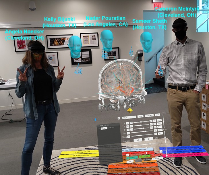
The challenge is compounded when expert surgeons need to collaborate. A neurosurgeon in North Carolina might need input from a colleague in Ohio. Traditional methods — sharing screen captures, describing anatomy over phone calls — fall far short of what these precision procedures demand.
The Solution: Life-Sized Holographic Planning
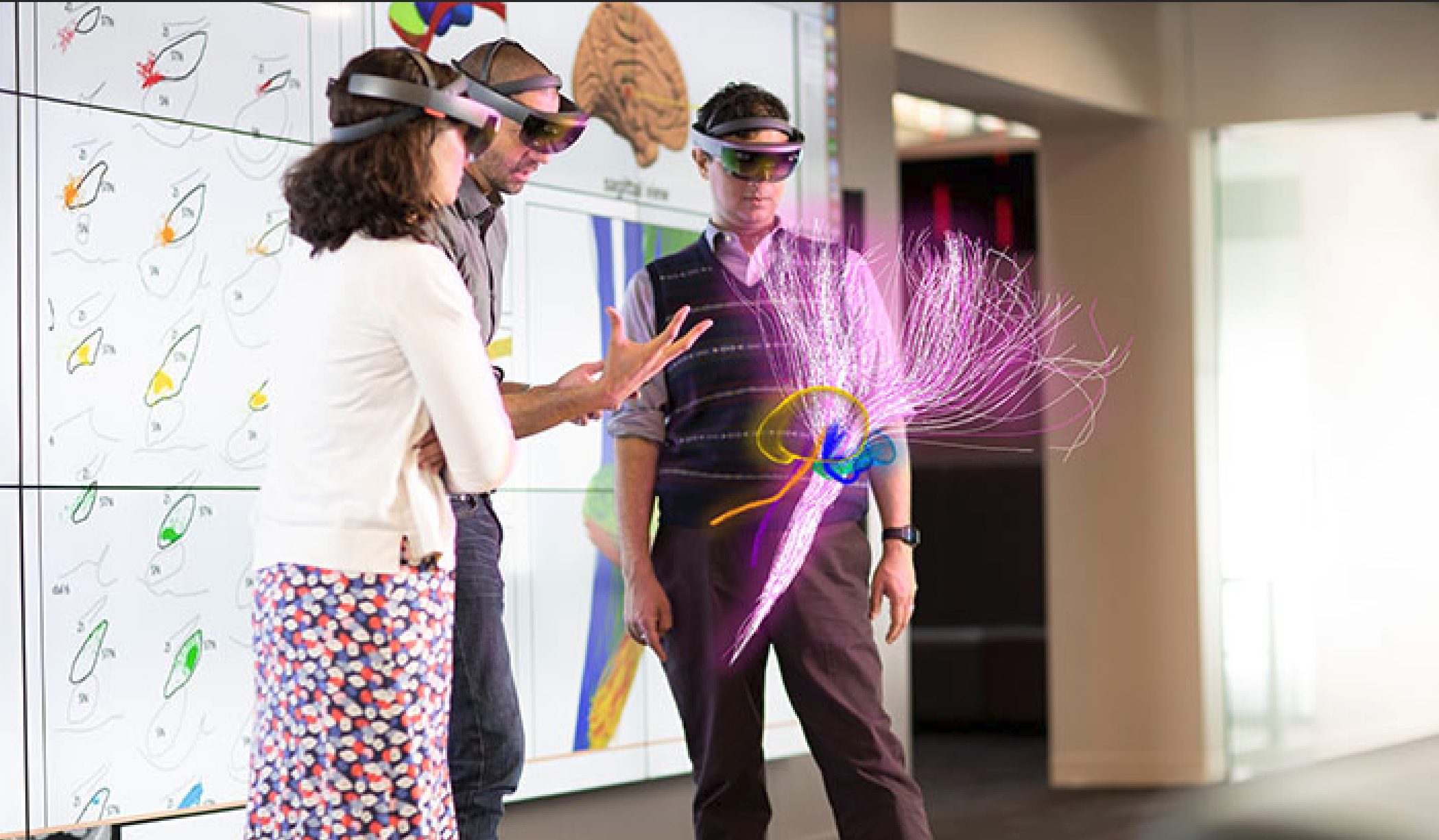
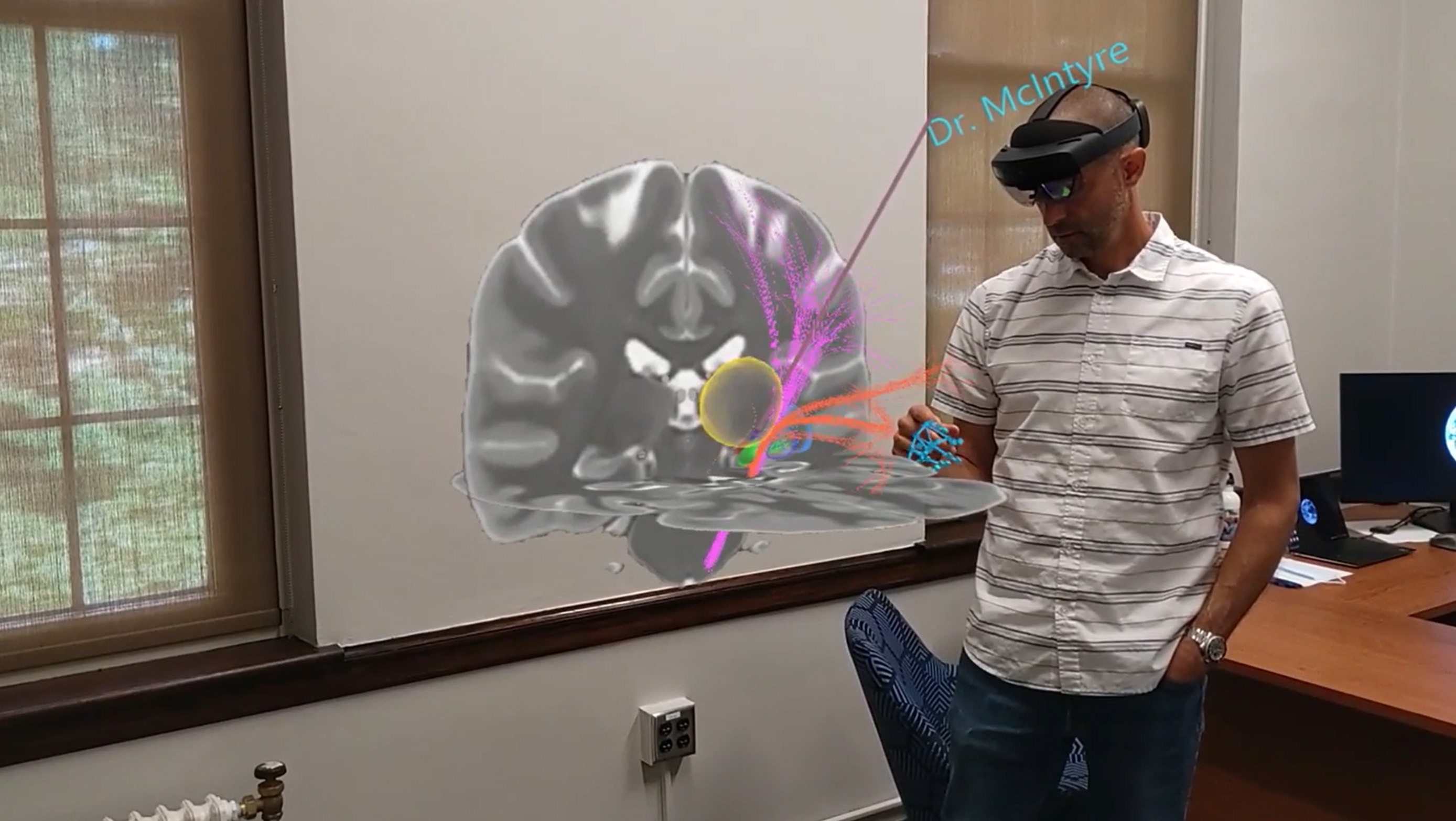
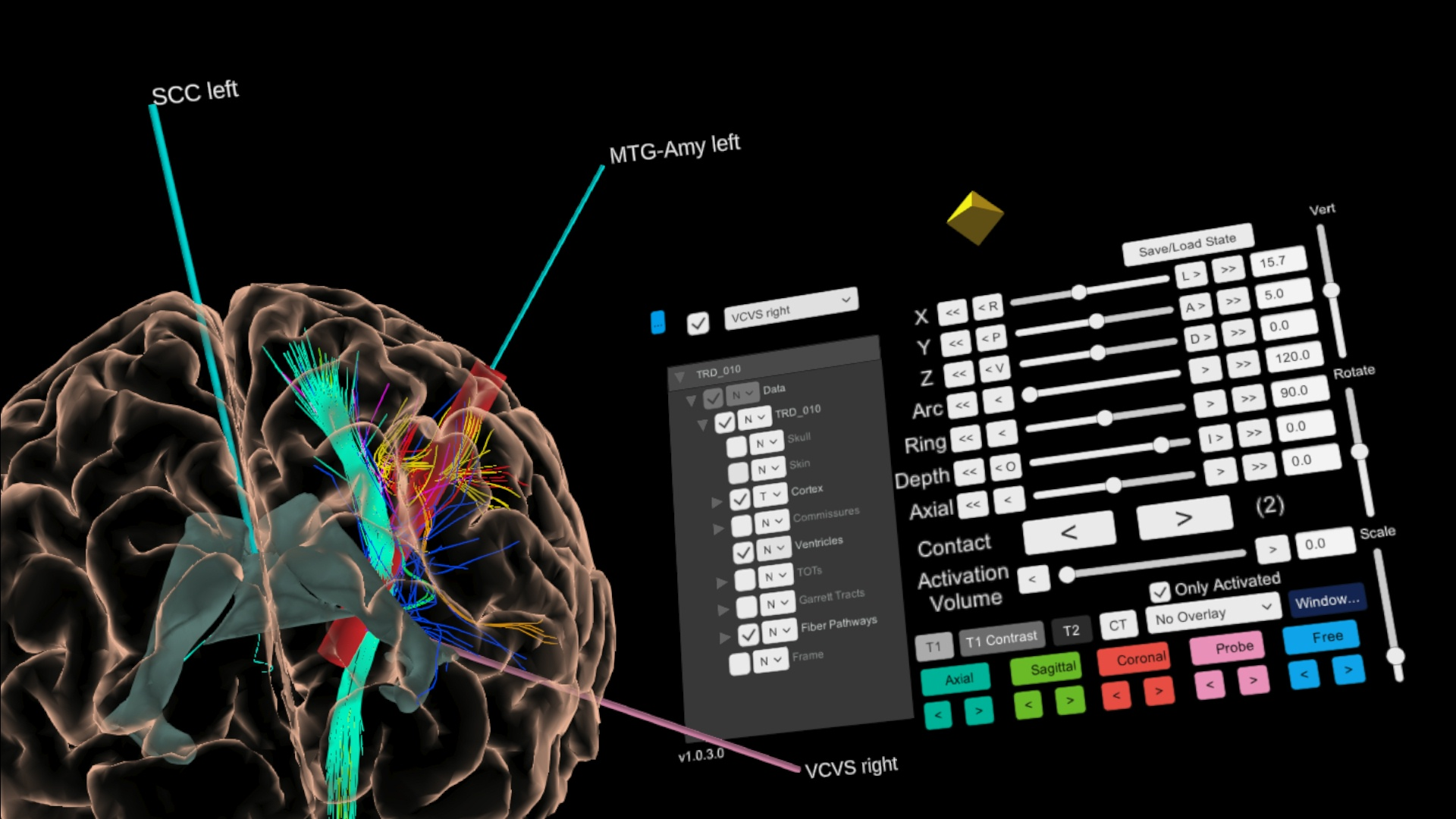
Using CrewXR, the surgical team examines the patient's brain scan as a life-sized hologram. Neural pathways — the delicate white matter tracts that connect regions of the brain — are rendered as glowing 3D streams using dynamic tractography. Surgeons can walk around the hologram, lean in to examine structures, and trace pathways with their hands.
Critically, CrewXR's real-time synchronization means a colleague 500 miles away sees the exact same hologram simultaneously. They can point to structures, annotate concerns, and collaboratively identify the precise coordinates for electrode placement. The immersive, spatial nature of the interaction reveals relationships between structures that are invisible in 2D views.
The Impact: From Research to the Operating Room
This work earned a $2.3 million NIH grant and was published in Neuron, one of the most prestigious journals in neuroscience. But the real measure of success is that this technology has been used in actual surgical operations — not as a demonstration, but as a tool that surgeons rely on to plan life-changing procedures.
The project produced the first-ever interactive holographic brain map, establishing a new paradigm for neurosurgical planning. It demonstrated that shared, immersive 3D visualization isn't just more engaging — it's fundamentally more effective for the kind of spatial reasoning that surgery demands.
“The ability to visualize and interact with neural pathways in three dimensions fundamentally changes how we plan these critical procedures.”